


Cards In This Set
| Front | Back |
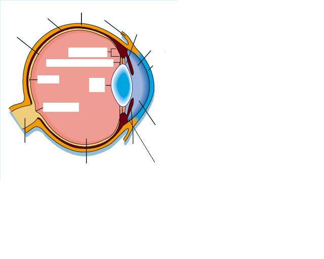 Label the Diagram |
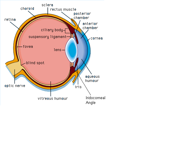 Answer |
|
What are the layers of the eye?
|
1. Fibrous tunic
2. Vascular tunica 3. Retina |
|
WHat makes up the fibrous tunica?
|
THe sclera and the cornea
|
|
Why is the cornea curved?
|
To focus light
|
|
Cornea histology?
|
Outer - non-keratinised stratified squamous epithelium
Middle - collagen and fibroblast - transparent due to the regular pattern of collagen fibres Inner - corneal endothelium |
|
How does the cornea get oxygen?
|
No blood supply so it diffuses from the air.
|
|
WHat is the role of the sclera?
|
It maintains shape and protects
|
|
What is the sclera made of?
|
Connective tissue and fibroblasts.
|
|
What is the conjunctiva?
|
It covers the visible and the part of the sclera under the eyelid.
|
|
What makes up the vascular tunica?
|
1. Choroid
2. Ciliary body 3. Iris |
|
Describe the choroid.
|
It is the highly vascular posterior portion of the vascular tunica. it has melanocytes that secrete melanin which absorbs reflected light to help produce sharp image.
|
|
Describe the ciliary body.
|
It is an anterior modification of the choroid with ciliary process which secrete aqueous fluid. It is also has ciliary muscles which are attached to the lens via zonular fibres which allow for accomodation
|
|
Describe the iris.
|
It is another choroid modification which lies between the lens and cornea. It contains melanocytes and circular and radial muscles.
|
|
What is the function of the iris?
|
When exposed to bright light the parasympathetic nerves cause contraction of the circular muscle which decreases the diameter of the pupil. When exposed to darkness the sympathetic stimulation stimulates the radial muscle which increases pupil diameter.
|
|
What are the two layer of the retina?
|
1.Pigmented layer - epithelium + melanocytes
2. Neuronal layer |
