


Cards In This Set
| Front | Back |
|
Benign Fibro-Osseous Lesions
|
- a collection of non-neoplastic intraosseous lesions that replace normal bone and consist of a cellular fibrous CT within which non-functional osseous structures form
- normal bone is replaced by fibrous CT and becomes ossified in a non-functional manner (first is radiolucent then becomes radiopaque) - 3 types - cemento-osseous lesions, fibrous dysplasia, cherubism |
|
Cemento-Osseous Lesions
|
- benign fibro-osseous lesions of the jaws closely associated with the apices of the teeth and containing amorphous spherical calcifications thought to resemble an aberrant form of cementum
- lesion usually formed without any signs or symptoms - these are localized to the jaws and start in the apical with 2 subtypes (localized, generalized) |
|
Periapical Cemental Dysplasia (Cementoma)
|
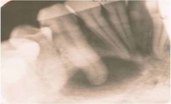 - this is asymptomatic diffuse periapical radiolucent and radiopaque areas, primarily in the anterior mandible - the cemento-osseous tissue replaces the normal bony archetecture - most commonly an African American female where there is a symptomless radiolucency at the apex of a tooth - this CANNOT be mistaken for an infection, clinical diagnosis |
|
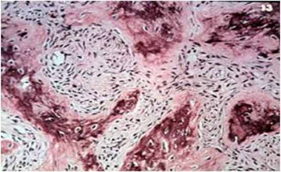
Histology of the Periapical Cemental Dysplasia
|
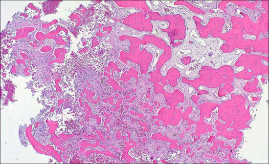 - note that this is usually present in the mandibular anterior but can be present in other regions as well - we do not need a biopsy to confirm this diagnosis, confirmed by clinical presentation and radiograph - the lesion is predisposed to infection and thus surgery will cause a possible unnecessary infection (possibly due to low vasculature) |
|
3 Phases of Periapical Cemental Dysplasia
|
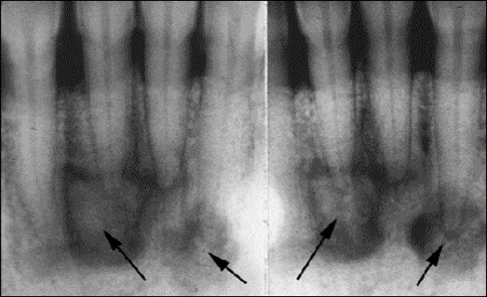 - 1) - osteolytic (radiolucent) - cellular CT replaces normal PA trabecular bone - 2) - cementoblastic (mixed RO & RL) where spherical calcifications fuse together and replace the CT - 3) - mature - mostly radiopaque - sclerotic mineralized bone with some CT interspersed within the mass |
|
Periapical Cemental Dysplasia Notes
|
- more common in women (90%)
- asymptomatic and are discovered accidentally - diagnosed clinically and radiographically (should not be biopsied) - no treatment is required |
|
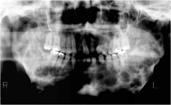
Florid Cemento-Osseous Dysplasia
|
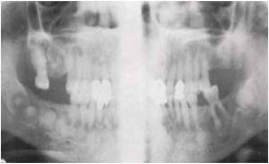 - diffuse asymptomatic radiopaque and radiolucent intraosseous areas of the cemento-osseous tissue that involves one or both arches (more generalized version of the periapical cemental dysplasia) - lesions have the same clinical presentation as the periapical cemental dysplasia, again patients are commonly African American and female |
|
Fibrous Dysplasia
|
 - this is similar to the periapical cemental dysplasia in that the bone is replaced with fibrous tissue and it becomes ossified - the difference is a gradual expansion of the bone and this is not unique to the bone - this is an asymptomatic regional alteration of the bone in which the normal architecture is replaced by fibrous tissue and non-functional trabeculae-like osseous structures - lesions may be monostotic or polystotic with or without associated endocrine disturbances |
|
Radiographic Pattern of Fibrous Dysplasia
|
 - a ground glass pattern is the type of radiographic change associated with this disease - also has been described as a cotton ball - histologically it resembles chinese characters which will become ossified |
|
Juvennile Fibrous Dysplasia
|
 - this is the most common form of the monostotic type of fibrous dysplasia of the head and neck - slow growing regional distortion that enlarges proportionately with the affected bone - an aggressive type of juvenile FD has a faster growth rate causing more functional difficulties for the patient |
|
Polystotic Fibrous Dysplasia
|
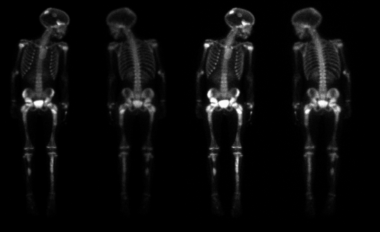 - this is fibrous dysplasia in many bones without any oter clinical findings, referred to as the jaffe type - patient with FD with functional disabilities, we wait until they mature and then can surgically recontour the affected bone |
|
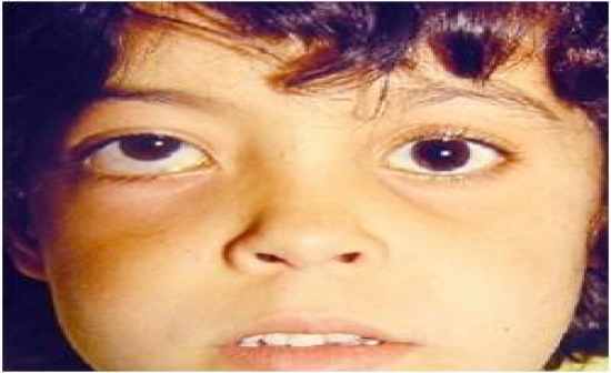
Albright Syndrome
|
- this is a form of polystotic fibrous dysplasia in many bones associated with a hormonal disorder, and brownish skin lesions (cafe au lait) - brown pigmentation of the skin
|
|
Adult Monostotic Fibrous Dysplasia
|
- this is a rare form of the fibrous dysplasia occuring spontaneously in adults
|
|
Treatment in Fibrous Dysplasia
|
- Surgery for establishment of function (speech, sight, breathing, mastication)
- surgery for cosmetic reasons after puberty in juvenile monostotic type |
|
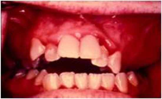
Cherubism (Bilateral Multilocular Radiolucencies)
|
 - autosomal dominant fibro-osseous lesion of the jaw involving more than one quadrant that stabilizes after the growth period, usually leaving some facial deformity and malocclusion - lesion of the jaw and no other region, expansile lesion of the jaw - occurs in males 2:1 males affected more severely - more common in the mandible than in the maxilla - biopsies produce an osteolytic lesion (fibrous lesion with multinucelated giant cells responsible for the radiographic appearance) |
