


Cards In This Set
| Front | Back |
|
Patients with visual object agnosia have no problem ________ing images. Their impairment comes in _____ing images.
|
Seeing, copying, and remembering
Recognizing, naming, and identifying
|
|
What brain region is associated with face recognition?
|
Fusiform gyrus/fusiform face area (FFA)
|
|
What do we mean when we say that visual object agnosia is a disorder of "associative" functioning but not "apperceptive" functions.
|
This means that the agnosia is not a visual problem, but rather involves an inability to associate images/objects with their semantic representations.
|
|
What are the three theories of what the fusiform face area (FFA) actually specializes in?
|
1. face recognition
2. global attribute recognition (arrangement of details within an image)
3. mediation of visual expertise (making fine distinctions based on learning)
|
|
Pure Alexia (difficulty reading words, exclusively) appears to result from lesions in the _________.
|
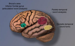 Visual Word Form Area |
|
Topographic amnesia involves an inability to __________ and results from lesions in the ____________.
|
Pictures of scenes
parahippocampal place area (ppa)
|
|
Object agnosia is thought to result from lesions in the _________.
|
Bilateral lingual-fusiform gyrus
|
|
In an early fMRI study by Malach, et al., what differentiated activity in the lateral occipital area (LO) from primary visual cortex (V1)?
|
LO was more active when subjects viewed pictures of objects, while V1 was more active during viewing of textures.
|
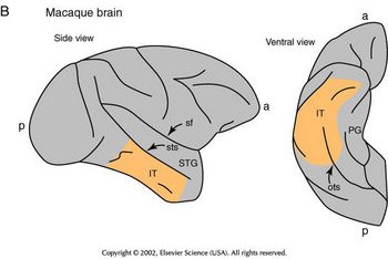 (Holmes &) Gross demonstrated visual object agnosia with lesions of inferior temporal cortex (IT). How did these monkeys compare with unoperated controls and striatal/foveal lesion monekys? |
Prior to surgery, all monkeys were trained to differentiate between two sideways letters. After surgery, IT lesion monkeys did not retain the ability to discriminate, while the other two groups did.
|
|
Desimone, et al. (Gross's lab) demonstrated neuronal selectivity for visual patterns in the monkey IT cortex. How?
|
Single-unit recordings of neurons while presenting certain objects (for ex, hands) and similar, more abstract shapes (human hands, moneky hands, rectangular fork shape, plain rectangle, etc.)
|
|
Desimone, et al. (Gross's lab) demonstrated that monkey IT cortex cells are sensitive to ________, but not to ________ or _______.
|
Patterns
motion, orientation
|
|
True or false? IT neurons respond more strongly to objects that are familiar to the monkey.
|
False. Familiarity is irrelevant.
|
|
Describe what Tanaka was trying to discover in the study where they presented 3d objects, then 2d objects, then simpler version of image.
|
They were trying to determine the minimal combination of features required to activate an IT neuron. ("subtractive analysis")
|
|
Describe, to the best of your ability, the disgustingly complicated experiment that Kobatake & Tanaka did, showing pictures of objects while recording from V2, V4, posterior IT, and anterior IT.
|
They compared the response of a neuron to its best simple stimulus (i.e., the best oriented colored bar) with its response to its best complex stimulus, calculating the Smax/Max ratio. From area V2 to anterior IT (back to front), there is a steady decrease in Smax/max ratio.
|
|
Using a very "subjective and arbitrary approach" (according to Carl) of just shoving a whole bunch of random objects in front of monkeys, Tanaka, et al (1991), found that, although responses of anterior IT cells were selective to particular complex features, _____________.
|
The features coded by individual cells were not complex enough to indicate a concept of particular real objects.
|
