


Cards In This Set
| Front | Back |
|
Glenoid Fossa
|
Under zygomatic/temporal connection towards posterior
Jaw doesn't go posteriorly unless trauma |
|
Lateral pterygoid muscle joins with
|
Articular disk
|
|
Ligaments of the TMJ
|
Articular capsule
|
|
Capsular ligament
|
Superiorly - temporal boneinferiorly - around neck of condyle contain synovial fluid Function: prevents anterior dislocation (limits down and forward)
|
|
Temporomandibular Ligament
|
OOP - Outer Oblique portionIHP - Inner horizontal portion Continuous with capsular ligamentattach- anteriorly to zygomatic process of temporal bone posteriorly to lateral side of condyle neck Function: main suspensory ligament during hinge movement. limits retraction of mandibule
|
|
Accessory ligaments
|
Sphenomandibularstylomandibular
|
|
Sphenomandibular ligament:
|
Originates: spine of sphenoid boneattaches: lingula of mandible function: suspensory ligament during wide opening when condyle moves forward becomes taut as temporomandibular loosens
|
|
Stylomandibular ligament
|
Origin: styloid processattach: posterior border of the ramus just above the angle function: limits anterior movement of angle during protrusion
|
|
Lateral and medial discal ligaments
|
Attach: medial and lateral poles of the condyle Divide the joint mediolaterally into the superior and inferior joint spaces
function: restrict movement of the disc away from the condyle |
|
Major planes of mandibular movement
|
Horizontal + sagittal
|
|
Articular tubercle
|
Anterior wall = articular eminence medial
|
|
Meniscus
|
Articular disc thinnest in the intermediate thickest in the posterior thicker medially than laterally devoid of blood vessels or nerve fibers attaches to capsular ligament joint cavity above and below disc- lining cells produce synovial fluid attaches distally to retrodiscal tissuesuperiorly: sup. retrodiscal lamina- to tympanic plate
|
|
TMJ anatomy
|
Long axis of head is oriented medio-posteriorly anterosuperior surface articulates with anterior wall of glenoid fossa sits on thinner intermediate zone of the disc
|
|
Posterior margin of articular surface of condyle attaches to
|
Inferior retrodiscal lamina
|
|
TMJ
|
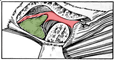 Pic |
