


Cards In This Set
| Front | Back |
 V |
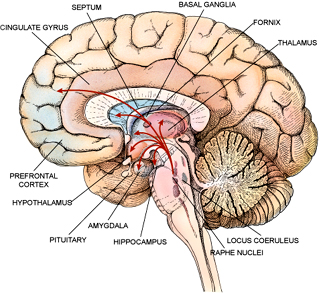
Mid-Sagittal Brain
|
|
A thin mantle of gray matter about the size of a formal dinner napkin covering the surface of each cerebral hemisphere. The cerebral cortex is crumpled and folded, forming numerous convolutions (gyri) and crevices (sulci). It is made up of six layers of nerve cells and the nerve pathways that connect them. The cerebral cortex is responsible for the processes of thought, perception and memory and serves as the seat of advanced motor function, social abilities, language, and problem solving.
|
Cerebral Cortex
|
|
Is a structure of the mammalian brain in the longitudinal fissure that connects the left and right cerebral hemispheres. It facilitates communication between the two hemispheres. It is the largest white matter structure in the brain, consisting of 200-250 millioncontralateral axonal projections. It is a wide, flat bundle of axons beneath the cortex. Much of the inter-hemispheric communication in the brain is conducted across the corpus callosum
|
Corpus Callosum
|
|
Is between the cerebral cortex and the midbrain, both in terms of its location and its neurological connections. Its function includes relaying sensation, special sense and motor signals to the cerebral cortex, along with the regulation of consciousness, sleep and alertness. The thalamus surrounds the third ventricle. It is the main product of the embryonic diencephalon.
|
Thalamus
|
|
The medulla oblongata is the lower half of the brainstem. In discussions of neurology and similar contexts where no ambiguity will result, it is often referred to as simply the medulla. The medulla contains the cardiac, respiratory, vomiting and vasomotor centers and deals withautonomic functions, such as breathing, heart rate and blood pressure.
|
Medulla
|
|
S a region of the brain that plays an important role in the integration of sensory perception, coordination and motor control. In order to coordinate motor control, there are many neural pathways linking the cerebellum with the cerebralmotor cortex (which sends information to the muscles causing them to move) and the spinocerebellar tract (which provides proprioceptivefeedback on the position of the body in space). The cerebellum integrates these pathways using the constant feedback to fine-tune motor activity.[1]
|
Cerebellum
|
|
1
|
2
|
|
3
|
4
|
|
Is a long, thin, tubular bundle of nervous tissue and support cells that extends from the brain.
|
Spinal Chord
|
|
Is an endocrine gland about the size of a pea. It is a protrusion off the bottom of the hypothalamus at the base of the brain, and rests in a small, bony cavity (sella turcica) covered by a dural fold (diaphragma sellae). The pituitary fossa, in which the pituitary gland sits, is situated in the sphenoid bone in the middle cranial fossa at the base of thebrain.The pituitary gland secretes hormones regulating homeostasis, including tropic hormones that stimulate other endocrine glands. It is functionally connected to the hypothalamus by the median eminence.
|
Pituitary
|
|
Is a portion of the brain that contains a number of small nuclei with a variety of functions. One of the most important functions of the hypothalamus is to link the nervous system to the endocrine system via the pituitary gland (hypophysis). is located below the thalamus, just above the brain stem. In the terminology of neuroanatomy, it forms the ventral part of the diencephalon. All vertebrate brains contain a hypothalamus. In humans, it is roughly the size of an almond. is responsible for certain metabolic processes and other activities of the Autonomic Nervous System. It synthesizes and secretes neurohormones, often called hypothalamic-releasing hormones, and these in turn stimulate or inhibit the secretion of pituitaryhormones. The hypothalamus controls body temperature, hunger, thirst, [1] fatigue, and circadian cycles.
|
Hypothalamus
|
|
is the part of the brain where the optic nerves (CN II) partially cross. The optic chiasm is located at the bottom of the brain immediately below the hypothalamus. Specifically, in the optic chiasm, the nerves connected to the right eye that attend to the left temporal visual field (located in the right retina) remain on the right (ipsilateral) side of the brain, and the nerves from the left eye that attend to the right temporal visual field (located in the left retina) remain on the left (ipsilateral) half of the brain
|
Optic chiasm
|
|
The Anterior Commissure (precommissure) is a bundle of nerve fibers (white matter), connecting the two cerebral hemispheres across the midline, and placed in front of the columns of the fornix. In a sagittal section, the anterior commissure is oval in shape, having a long vertical axis that measures about 5 mm.
|
Anterior Commissure
|
|
Is a rounded band of white fibers crossing the middle line on the dorsal aspect of the upper end of the cerebral aqueduct. It is important in the bilateral pupillary light reflex.
|
Posterior Commissure
|
|
S a small endocrine gland in the vertebrate brain. It produces melatonin, a hormone that affects the modulation of wake/sleep patterns and photoperiodic (seasonal) functions.[1][2] It is shaped like a tiny pine cone (hence its name), and is located near to the center of the brain, between the twohemispheres, tucked in a groove where the two rounded thalamic bodies join.
|
Pineal Body
|
