


Cards In This Set
| Front | Back |
|
HOLTER MACHINE
|
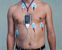 Is a portable device for continuously monitoring various electrical activity of the heart for at least 24 hrs often for 2 weeks at a time. |
|
ECG ELECTROCARDIOGRAM
|
 Is the process of recording electrical activity of the heart. |
|
ECG PAPER
|
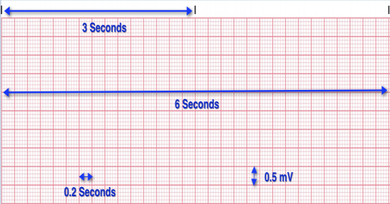 ECG is recorded on grid paper. The horizontal axis record time, with black marks at the top indicating 3sec. The vertical axis records amplitude (voltage). Small block = 0.1mV. |
|
ECG PAPER
|
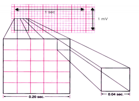 Every paper is marked by 5 large grid blocks. Each large grid block = 0.20sec. Within the large blocks there are 5 small blocks, each representing 0.04 sec. Speed 25mm/sec. |
|
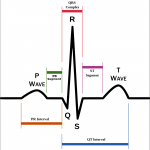
P WAVE
|
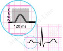 Represents atrial depolarization (the time necessary for an electrical impulse from the SA node to spread throughout the atrial musculature). Amplitude 2 to 2.5 mm, Duration 0.06 to 0.11 sec |
|
P-R INTERVAL
|
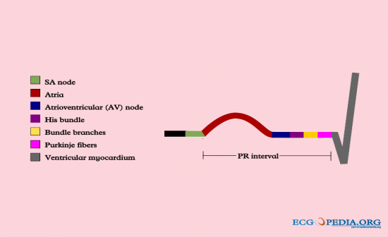 Represents the time it takes an impulse to travel from the atria to AV node, bundle of His, bundle of branches to the Purkinje fibers. Goes from the beginning of P wave to the beginning of QRS. Duration 0.12 to 0.20 sec. |
|
QRS COMPLEX
|
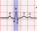 Represents ventricular depolarization, consists of 3 waves the Q wave, the R wave and the S wave. Q wave is not always present. R is a positive deflection and S is a negative deflection. Follows the P-R interval, amplitude varies with age and sex and the duration is 0.06 to 0.12 sec. (1.5 to 3 small blocks) |
|
QT INTERVAL
|
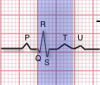 Represents the time necessary for ventricular depolarization and repolarization. Measure to the beginning of QRS complex to the end of T wave. duration varies according to age, sex and HR, 0.36 to 0.44sec (9-11 small blocks) |
|
T WAVE
|
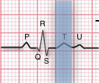 Indicates the repolarization of the ventricles, is an slightly asymmetrical and follows the QRS complex after a pause. Rare times U wave follows T wave. The U wave represents the repolarization of the Bundle of His and Purkinje fibers. Amplitude 5mm or less in standard leads I, II, III; 10mm or less in V1-V6. not measured. |
|
ST SEGMENT
|
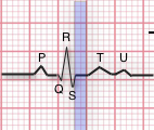 Represents the end of ventricular depolarization and the beginning of repolarization. Extends from the end of the S wave to the beginning of the T wave. Not masured |
|
THE 6 SECOND RULE TO DETERMINE HEART RATE
|
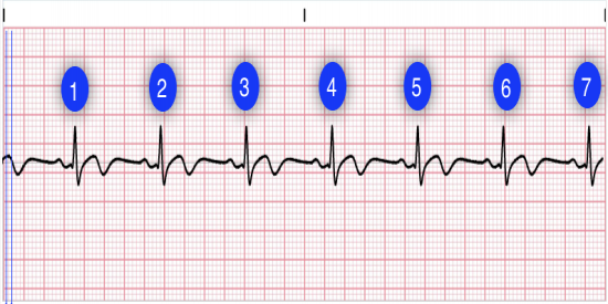 Count the number of QRS complexes over a 6 second interval. Multiply by 10 to determine heart rate. This method works well for both regular and irregular rhythms. In the first image, we can count 7 QRS complexes, so the heart rate is 70. |
|
DETERMINE HR 2ND METHOD
|
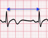 Count the number of small boxes for a typical R-R interval. Divide this number into 1500 to determine heart rate. In the second image, the number of small boxes for the R-R interval is 22.5. The heart rate is 1500/21.5, which is 69.8. (70BPM) |
|
CHEST LEADS VIEW WHAT PART OF THE HEART
|
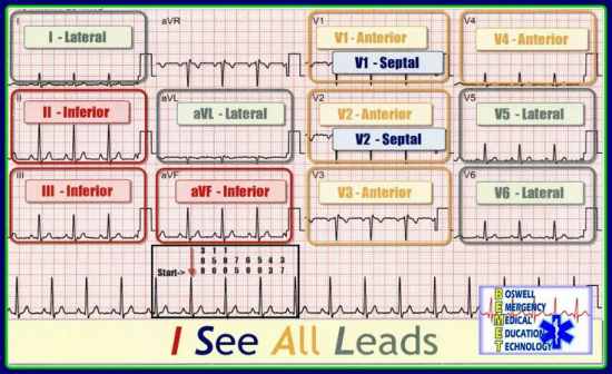
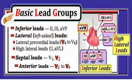 V1 & v2 (Right Ventricle)V3 & V4 (septum/lateral left ventricleV5 & V6 (Anterior/Lateral Left Ventricle) |
|
CHEST ELECTRODE PLACEMENT
|
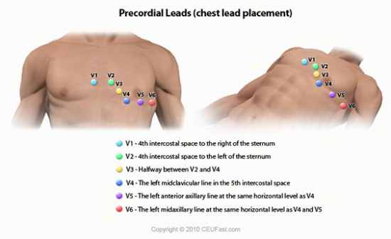 V1: Fourth intercostal space to the right of the sternum. V2: Fourth intercostal space to the Left of the sternum. V3: Directly between leads V2 and V4. V4: Fifth intercostal space at midclavicular line. V5: 5th ICS at left anterior axillary line. V6: 5th ICS at left midaxillary line. (Directly under the midpoint of the armpit) |
|
LEAD GROUPS
|
 |
