MCAT Biology Ch. 11 The Musculoskeletal System

54 cards   |   Total Attempts: 182
  

Cards In This Set

Front Back
3 Main Types of Muscle
  1. Skeletal
  2. Smooth
  3. Cardiac
Skeletal Muscle
  • Functions:
    • Support & movement
    • Propulsion of blood thru veins
    • Thermoregulation (shivering)
  • Characteristics:
    • Multinucleated
    • Striated
    • Under voluntary (somatic) control
    • Divided into 2 types of fibers:
      • Red (Slow-Twitch) Fibers - oxidative phosphorylation
      • White (Fast-Twitch) Fibers - anaerobic metabolism
Smooth Muscle
  • Located in respiratory, reproductive, cardiovascular, & digestive systems
  • Characteristics:
    • Nonstriated
    • Under involuntary (autonomic) control
    • Uninucleated (only 1 nucleus)
    • * Myogenic Activity - contraction w/out any neural input
Cardiac Muscle
  • Comprises contractile tissue of the heart
  • Characteristics:
    • Striated
    • Under involuntary (autonomic) control
    • Can be uni- or binucleated (1 or 2 nuclei)
    • Cells are connected w/ intercalated discs that contain gap junctions
    • * Myogenic Activity - contraction w/out any neural input
Red Fibers (Slow-Twitch Fibers)
  • High myoglobin content (red color)
  • Derive energy aerobically
  • Many mitochondria for oxidative phosphorylation
White Fibers (Fast-Twitch Fibers)
  • Contain much less myoglobin than red fibers (white color)
  • Derive energy mostly anaerobically
Intercalated Discs (& Gap Junctions)
Cardiac muscle cells are connected by intercalated discs which contain many gap junctions. Gap junctions are connections between the cytoplasm of adjacent cells, allowing for the flow of ions directly between cells. Allows for rapid & coordinated muscle cell depolarization & efficient contraction of cardiac muscle
Sarcomere
Basic contractile unit of striated (skeletal) muscle.
  • Made of thick (myosin) & thin (actin) filaments
Troponin & Tropomyosin
Found on the thin filament & regulate actin-myosin interactions
Myofibrils
Sarcomeres attach end to end to become myofibrils
Lines, Zones, & Bands of the Sacromere
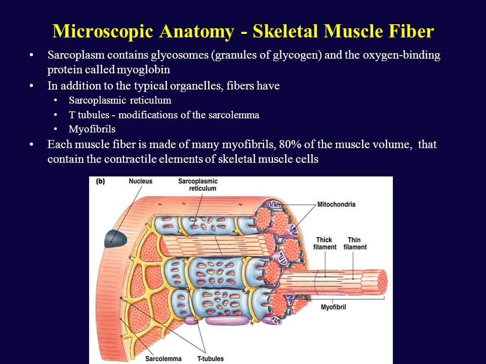
M-Line - located in middle of sarcomereI-Band - contains only thin filamentsH-Zone - consists of only thick filamentsA-Band - contains thick filaments in their entirety, including any overlapping area w/ thin filaments. **Only part of sacromere that maintains constant size during contraction!!!
  • M-Line - located in middle of sarcomere
  • I-Band - contains only thin filaments
  • H-Zone - consists of only thick filaments
  • A-Band - contains thick filaments in their entirety, including any overlapping area w/ thin filaments. **Only part of sacromere that maintains constant size during contraction!!!
M-Line - located in middle of sarcomereI-Band - contains only thin filamentsH-Zone - consists of only thick filamentsA-Band - contains thick filaments in their entirety, including any overlapping area w/ thin filaments. **Only part of sacromere that maintains constant size during contraction!!!
Myocytes
A muscle cells or muscle fiber. Made up of many myofibrils
Sarcoplasmic Reticulum
Surrounds myofibrils, is a calcium-containing modified endoplasmic reticulum
Sarcolemma
Cell membrane of a myocyte
T-Tubules
A system of T-tubules is connected to the sarcolemma & oriented perpendicularly to the myofibrils, allowing incoming signals from nerves to reach all parts of the muscleA system of T-tubules is connected to the sarcolemma & oriented perpendicularly to the myofibrils, allowing incoming signals from nerves to reach all parts of the muscle