


Cards In This Set
| Front | Back |
|
List and describe the 6 levels of protection for the brain against mechanical trauma.
|
1. skin
2. cranium (and vertebral column) 3. dura mater (hard mother, thick, tough, inelastic. 2 layers - periosteal (attached to inner skull) and meningeal (attached to arachnoid) spinal dura has 1 layer only which attaches to coccynx at filium terminale externum.) 4. arachnoid (thin cell layer with arachnoid trabeculae - web like collagen projections into pia layer. CSF produced in choroid plexus flows in the sub-arachnoid space, presence of subarachnoid cisterns). 5. pia mater (tender mother, thin, delicate, tightly covering all contours, highly vascular. spinal pia layer has specialisations - denticulate ligaments attaching lateral cord to arachnoid, filium terminale internum). arachnoid + pia = leptomeninges 6. CSF (buoyancy, cushioning, removes waste products, produced by choroid plexi) |
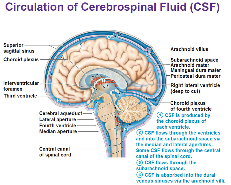 Draw a diagram and describe the flow of CSF in and around the brain. Be as specific as possible. |
*CSF is produced by the CP in each of the ventricles. the majority originates from the CP in the lateral ventricles.
*flows through intraventricular foramen into the third ventricle. *CP located on roof of third ventricle adds more CSF. *fluid then moves through the cerebral aqueduct within the midbrain and passes into the rhomboid-shaped fourth ventricle. *CP of fourth ventricle adds more CSF. *CSF leaves ventricular system through the median and lateral apertures (aka foramen of magendie/foramen of luschka) of fourth ventricle and enters subarachnoid space. *from here it may flow over the cerebral convexities or into the spinal subarachnoid space. |
|
What is a sub-arachnoid cistern?
Name 4 subarachnoid cisterns. |
The areas in which the subarachnoid and pia maters are widely separated
from each other leading to the widening of the subarachnoid space
1. cisterna magna 2. pontine cistern 3. interpeduncular cistern 4. quadrigeminal cistern |
|
What are arachnoid villi?
Describe their role in waste removal |
Arachnoid villi are small protrusions of the arachnoid through the dura mater.
they protrude into the venous sinuses of the brain, and allow cerebrospinal fluid (CSF) to exit the sub-arachnoid space and enter the blood stream. the arachnoid villi act as one-way valves. normally the pressure of the CSF is higher than that of the venous system, so CSF flows out through the villi and into the blood. |
|
Why does the brain need protection from blood?
|
Because neuron function depends on:
*specific EC ion concentrations *NT-receptor binding *EC growth factors important to neurons and glia blood composition is constantly changing in ion concentration, hormones, glucose, nutrients and oxygen. in comparison the brain's CNS environment is far more refined, and must be protected from the chemical changes associated with blood. |
|
Describe how the true blood brain barrier is formed
|
*by the formation of tight junctions between endothelial cells lining the capillaries.
*these tight junctions are induced between capillary endothelial cells by astrocytes present during development. i.e. in the absence of astrocytes, tight junctions do not form. |
|
If we have a barrier protecting the brain from blood, how does the brain get the required nutrition from the blood? Give examples
|
*gases diffuse across cell membrane
*lipid soluble substances cross endothelial cells through membranes (e.g. alcohol, steroid hormones) i.e. greater lipid solubility = easier entry *via carrier molecules (only selected essential substances e.g. glucose, large aa's like phe. these are synthesised by capillary endothelial cells and are a form of active transport - thus capillary endothelial cells are full of mitochondria for ATP). |
|
What substances can and cannot cross the true blood-brain barrier and why. Give specific examples.
|
Can: small hydrophobic molecules like gases and hormones (diffusion) , lipid soluble substances e.g. valium, nicotine, chloramphenicol; selected substances e.g. glucose (energy), large aa's e.g. phe --> precursor for NT synthesis.
can't: most water-soluble substances (e.g. penicillin, L-glucose), bacteria |
|
What is phenylketonuria? How does this occur?
|
Phenylketonuria is an autosomal recessive metabolic genetic disorder characterized by a mutation in the gene for the hepatic enzyme phenylalanine hydroxylase (PAH), rendering it nonfunctional. This enzyme is necessary to metabolize the amino acid phenylalanine (Phe) to the amino acid tyrosine. When PAH activity is reduced, phenylalanine accumulates causing toxicity (-->brain damage) and decreased tyrosine (a NT precursor - dopamine).
|
|
How does the choroid plexus form a barrier between the blood and CSF?
|
*CP is 3 layered (choroid epithelial cells, pia mater, capillaries)
*the choroid capillaries lie outside the pia and are fenestrated *but the choroid epithelial cells have tight junctions and prevent flow of blood-borne components into the CSF |
|
What is the function of the choroid plexus?
|
*produce CSF
*provide 2-way selective flow from blood to the CSF -selected substances actively transported across the choroid epithelial cells (e.g. Na, Cl, K, C & B vitamins --> water soluble) -choroid epithelial cells have extensive microvilli on their ventricular surface increasing surface area for waste removal from CSF to blood |
|
What is hydrocephaly? Describe 3 instances in which this can occur.
|
*hydrocephaly is when too much CSF is produced and accumulates within the ventricles and cavities of the brain
*happens due to tumors, poor removal or obstructed removal of CSF |
|
What is the arachnoid barrier?
|
*barrier that prevents the transfer from extra-cerebral capillaries into the CSF in the sub-arachnoid space
*it is formed by tight junctions of the arachnoid cells |
|
List the arteries that make up the circle of Willis.
What is the significance of having these arteries arranged in a circular manner? |
.internal carotids
.cerebral group (anterior, middle, posterior cerebral arteries) .communicating arteries (anterior, posterior) the significance of the arrangement of the arteries allows the compensation for small blockages within the circle. |
|
Name the 2 main types of stroke and give examples of how they can occur.
|
1. ischaemic: happens when a vessel is blocked or closed and cuts of blood supply to an area of the brain. e.g. thrombus (blot clot within the vessel), embolism (foreign material carried in the blood).
2. hemorrhagic: leakage of blood flow into brain tissue. e.g. ruptured aneurysm, arteriovenous malformation (AVM - a malformation prone to hemorrhage), weakened vessel walls due to hypertension |
